Stress Fractures
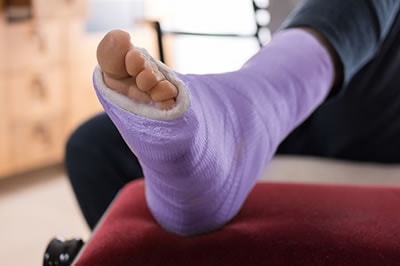 Stress fractures are incomplete cracks in the bone, generally in the foot; the metatarsal bones are most affected. This can occur in any age group. Many times this will occur in children or adults who begin a new activity after a period of inactivity. Pain and swelling are noted on the top of the instep, and there is increased pain with toe-off or pushing off as you walk. These fractures can be treated with complete rest. However, at North Bay Foot & Ankle Center, we maintain activity during our rehabilitation program. We find that our patients do better to maintain activity while healing, and this eliminates the need to rehabilitate the adjoining structures.
Another name for stress fracture is March fracture. This was coined in the service when the recruits began their training, which consisted of marching. The repeated trauma caused the fractures. Most recruits marched on through their injuries and graduated from basic training with the bone well healed. Most times the bone is stronger after healing from this type of fracture. Certain foot types are prone to stress fractures and many times with this type of foot, a second stress fracture may occur in the adjacent bones after the first fracture heals. This is nature's way of realigning the weight distribution.
Foot surgeons have designed procedures, which raise or lower metatarsals depending on the symptoms based on what they observed in these healing stress fractures.
Stress fractures are sometimes misdiagnosed as more severe problems to those unfamiliar with the callous formation, which occurs while the fracture heals. Many times it is only when the callous occurs on the x-ray, 10 to 14 days after injury, that we actually see the fracture on film. Remember these are round bones and the x-rays are two-dimensional.
Treatment of these problems at North Bay Foot & Ankle consist of diagnosis, rehabilitation, and prevention. Diagnosis by clinical evaluation and x-ray, rehabilitation by flexible strapping, H-wave therapy, ultrasound, and prevention with certain orthotics.